Structural Biochemistry/Collagen
Collagen Introduction
Collagen, which is the most abundant protein in mammals, is also the main fibrous component of skin, bone, tendon, cartilage, and teeth. Humans' dry weight of skin are made up of over 1/3 collagen. This extracellular protein is a rod-shaped molecule, about 3000 Å long and only 15 Å in diameter. There are at least twenty-eight different types of collagen that are made up of at least 46 different polypeptide chains that have been located in vertebrae and other proteins that contain collagenous domains. The defining characteristic of collagen is that it is a structural proteins that are composed of a right handed bundle of three parallel-left handed polyproline II-type helices. Because of the tight packing of PPII helices within the triple helix, every third residue, which is an amino acid, is Gly (Glycine). This results in a repeating pattern of an XaaYaaGly sequence. Although this pattern occurs in all types of collagen, there is some disruption of this pattern in certain areas located in within the triple helical domain of nonfibrillar collagens. The amino acid that replace the Xaa in the sequence is most likely (2S) –proline (Pro, 28%). The most likely replacement amino acid in the Yaa position is (2s,4R)- 4-hydroxyproline (Hyp, 38%). This means that the ProHypGly sequence is the most common triplet in collagen. Many research has been done on figuring out the structure of the collagen triple helices and how their chemical properties affects collagen's stability. It has been found that stereo electronic effects and preorganization are important factors in determining the stability of collagen. A type of collagen called type I collagen has the structure revealed in detail. Synthesizing artificial collagen fibrils, which are smaller strands of fiber, have now been possible and can now contain properties that natural collagen fibrils have. By continually understanding the mechanical and structural properties of native collagen fibrils, will help research devise and develop ways to create artificial collagenous materials that can be applied to many aspects of our lives such as biomedicine and nanotechnology.

Structure of Collagen
The structure of collagen has been developed intensively throughout history. At first, Astbury and Bell put forth their idea that collagen was made up a single extended polypeptide chain with all their amide bonds in the cis conformation. In 1951, other researches correctly determined the structures for alpha helix and the beta sheet. Pauling and Corey put forth their structure that three polypeptide strands are formed together through hydrogen bonds in a helical conformation. In 1964, Ramachandran and Kartha developed an advanced structure for collagen in that it was a right handed triple helix of three left handed polypeptide 2 helices with all the peptide bonds in the trans conformation and two hydrogen bonds in each triplet. Afterwards, the structure was honed by Rich and Crick to the accepted triple helix structure today, which contains a single interstrand N-H(Gly)...O=C(Xaa) hydrogen bond per triplet and a tenfold helical symmetry with a 28.6 A axial repeat.
Function and diversity
Collagen, which is present in all multicellular organism, is not one protein but a family of structurally related proteins. The different collagen proteins have very diverse functions. The extremely hard structures of bone and teeth contain collagen and a calcium phosphate polymer. In tendons, collagen forms rope-like fibers of high tensile strength, while in the skin collagen forms loosely woven fibers that can expand in all directions. The different types of collagen are characterized by different polypeptide compositions. Each collagen is composed of three polypeptide chains, which may be all identical or may be of two different chains. A single molecule of type I collagen has a molecular mass of 285kDa, a width of 1.5nm and a length of 300nm.
| Type | Polypeptide Composition | Distribution |
|---|---|---|
| I | [alpha 1(I)]2, alpha 2(I) | Skin,bone,tendon,cornea,blood vessels |
| II | [alpha 1(II)]3 | Cartilage, intervertebral disk |
| III | [alpha 1(III)]3 | Fetal skin,blood vessels |
| IV | [alpha 1(IV)]2, alpha 2(IV) | Basement membrane |
| V | [alpha 1(V)]2, alpha 2(V) | Placenta,skin |
Overview of Biosynthesis
Collagen polypeptides are synthesized by ribosomes on the rough endoplasmic reticulum (RER). The polypeptide chain then passes through the RER and Golgi apparatus before being secreted. Along the way it is post-translationally modified: Pro and Lys residues are hydroxylated and carbohydrate is added. Before secretion, three polypeptide chains come together to form a triple-helical structure known as procollagen. The procollagen is then secreted into the extracellular spaces of the connective tissue where extensions of the polypeptide chains at both the N and C termini (extension peptides) are removed by peptidases to form troppcollagen. The tropocollagen molecules aggregate and are extensively cross-linked to procuce the mature collagen fiber.
Stability of Triple Helix Structure
Collagen is important for animals as it contains many essential properties such as thermal stability, mechanical strength, and the ability to bond and interact with other molecules. Knowing how these properties are affected require an understanding of the structure and stability of collagen. Replacing amino acids in place of any of the XaaYaaGly positions can affect the structure and stability of collagen in numerous ways.
Glycine Substitutions
Replacing the Glycine position in the XaaYaaGly sequence often cause diseases has it is associated with mutations in the triple helical and non triple-helical domains of a variety of collagens. The damaging mutations to collagen is caused by the substitution of Gly involved in the last hydrogen bods within the triple helix. For example the amino acid replacing the Gly and the location of the substitution can effect the pathology of osteogenesis. Substituting the Gly in proline rich areas of the collagen sequence have less disruption then the areas of proline poor regions. The time delay caused by Glycine substitutions results in an overmodification of the protocollagen chains, which alter the normal state of the triple helix structure and thus contributing the development of osteogenesis.
Higher-Order collagen Structure.
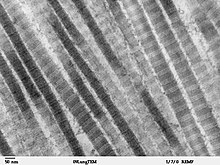
Collagen is made up hieracharcal components from the smaller units of individual TC monomers that self assemble into the macromolecular fibers. In type 1 collagen, monomers make up microfibrils which then make up fibris.
Fibril Structure.
TC monomers of type 1 collagen have a strange feature in that they are unstable at body temperature meaning they prefer to be disordered rather than structured and order. The question is that how can something unstable be a component of something so stable, like the triple helix structure of collagen. The answer to this question is that collagen fibrillogenesis stabilizes the triple helix, meaning when the monomers form together they have a stabilizing effect. This contributes to the strength of the collagen triple helix structure.
Collagen fibrillogenesis occurs through the formation of intermediate-sized fibril segments called microfibrils. There are two essential questions that need to be answered in order to understand the molecular structure of collagen fibrils. The first question is what is the arrangement of the individual TC monomers that make up the microfibril. The second question is then how do those microfibrils make up the collagen fibril. These questions are difficult to answer because individual natural microfibrils cannot be isolated and the big size and insolubility of mature collagen fibrils make it impossible for standard techniques to figure the structure out.


References
edit- ↑ Matthew D. Shoulders and Ronald T. Raines(2009). Collagen Structure and Stability'. "PubMed", p. 3-6.
- David Hames, Nigel Hooper. Biochemistry. 3rd edition. Taylor & Fancis Group, New York, 2005.
- Saad Mohamed (1994) "Low resolution structure and packing investigations of collagen crystalline domains in tendon using Synchrotron Radiation X-rays, Structure factors determination, evaluation of Isomorphous Replacement methods and other modeling." PhD Thesis, Université Joseph Fourier Grenoble 1 Link