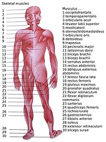
Science: An Elementary Teacher’s Guide/The Human Body: Muscles
The muscular system is an organ system consisting of skeletal, smooth and cardiac muscles. It permits movement of the body, maintains posture, and circulates blood throughout the body. The muscular system in vertebrates is controlled through the nervous system, although some muscles (such as the cardiac muscle) can be completely autonomous. Together with the skeletal system it forms the musculoskeletal system, which is responsible for movement of the human body.


Muscles
edit
a band or bundle of fibrous tissue in a human or animal body that has the ability to contract, producing movement in or maintaining the position of parts of the body.
There are three distinct types of muscles: skeletal muscles, cardiac or heart muscles, and smooth (non-striated) muscles. Muscles provide strength, balance, posture, movement and heat for the body to keep warm.
Upon stimulation by an action potential, skeletal muscles perform a coordinated contraction by shortening each sarcomere. The best proposed model for understanding contraction is the sliding filament model of muscle contraction. Actin and myosin fibers overlap in a contractile motion towards each other. Myosin filaments have club-shaped heads that project toward the actin filaments.
Larger structures along the myosin filament called myosin heads are used to provide attachment points on binding sites for the actin filaments. The myosin heads move in a coordinated style, they swivel toward the center of the sarcomere, detach and then reattach to the nearest active site of the actin filament. This is called a rachet type drive system. This process consumes large amounts of adenosine triphosphate (ATP).
Energy for this comes from ATP, the energy source of the cell. ATP binds to the cross bridges between myosin heads and actin filaments. The release of energy powers the swiveling of the myosin head. Muscles store little ATP and so must continuously recycle the discharged adenosine diphosphate molecule (ADP) into ATP rapidly. Muscle tissue also contains a stored supply of a fast acting recharge chemical, creatine phosphate which can assist initially producing the rapid regeneration of ADP into ATP.
Calcium ions are required for each cycle of the sarcomere. Calcium is released from the sarcoplasmic reticulum into the sarcomere when a muscle is stimulated to contract. This calcium uncovers the actin binding sites. When the muscle no longer needs to contract, the calcium ions are pumped from the sarcomere and back into storage in the sarcoplasmic reticulum.
Aerobic and anaerobic muscle activity
editAerobic exercise refers to endurance exercises such as biking, running or swimming. Anaerobic exercise refers to strength training and weight training. The difference is significant because aerobic and anaerobic muscle activity are fueled by different forms of energy, and have significantly different characteristics.
At rest, the body produces the majority of its ATP aerobically in the mitochondria without producing lactic acid or other fatiguing byproducts. During exercise, the method of ATP production varies depending on the fitness of the individual as well as the duration, and intensity of exercise. At lower activity levels, when exercise continues for a long duration (several minutes or longer), energy is produced aerobically by combining oxygen with carbohydrates and fats stored in the body. Activity that is higher in intensity, with possible duration decreasing as intensity increases, ATP production can switch to anaerobic pathways, such as the use of the creatine phosphate and the phosphagen system or anaerobic glycolysis. Aerobic ATP production is biochemically much slower and can only be used for long-duration, low intensity exercise, but produces no fatiguing waste products that can not be removed immediately from sarcomere and body and results in a much greater number of ATP molecules per fat or carbohydrate molecule. Aerobic training allows the oxygen delivery system to be more efficient, allowing aerobic metabolism to begin quicker. Anaerobic ATP production produces ATP much faster and allows near-maximal intensity exercise, but also produces significant amounts of lactic acid which render high intensity exercise unsustainable for greater than several minutes. The phosphagen system is also anaerobic, allows for the highest levels of exercise intensity, but intramuscular stores of phosphocreatine are very limited and can only provide energy for exercises lasting up to ten seconds. Recovery is very quick, with full creatine stores regenerated within five minutes.
Skeletal Muscles
edita muscle that is connected to the skeleton to form part of the mechanical system that moves the limbs and other parts of the body.
It is a form of striated muscle tissue which is under the voluntary control of the somatic nervous system. Most skeletal muscles are attached to bones by bundles of collagen fibers known as tendons.
A skeletal muscle refers to multiple bundles of cells called muscle fibers (fascicles). The fibres and muscles are surrounded by connective tissue layers called fasciae. Muscle fibres, or muscle cells, are formed from the fusion of developmental myoblasts in a process known as myogenesis. Muscle fibres are cylindrical, and have more than one nucleus.
Muscle fibers are in turn composed of myofibrils. The myofibrils are composed of actin and myosin filaments, repeated in units called sarcomeres, which are the basic functional units of the muscle fiber. The sarcomere is responsible for the striated appearance of skeletal muscle, and forms the basic machinery necessary for muscle contraction.
Cardiac Muscles
editCardiac muscle is heart muscle. Our heart is a muscle. It is a striated involuntary muscle.The cells that make up this muscle are called cardiomyocytes or myocardiocytes. The myocardium is the muscle tissue of the heart, and forms a thick middle layer between the outer epicardium layer and the inner endocardium layer. Cardiac muscle is the only muscle in the heart and no where else on the body. It's main job is to pump blood throughout the body.
Coordinated contractions of cardiac muscle cells in the heart propel blood out of the atria and ventricles to the blood vessels of the left/body/systemic and right/lungs/pulmonary circulatory systems. This complex mechanism illustrates systole of the heart.
Cardiac muscle cells, unlike most other tissues in the body, rely on an available blood and electrical supply to deliver oxygen and nutrients and remove waste products such as carbon dioxide. The coronary arteries help fulfill this function.
Smooth Muscles
editAre involuntary non-straited muscle. This muscle is found in the walls of organs such as oesophagus, stomach, bronchi, as well as the walls of blood vessels, lymphatic vessels, the urinary bladder, uterus, male and female reproductive tracts, gastrointestinal tract, respiratory tract, arrector pili of skin, the ciliary muscle, and iris of the eye. This muscle has slow rhythmic contractions that are stimulated by involuntary neurogenic impulses. The structure and function is basically the same in smooth muscle cells in different organs, but the inducing stimuli differ substantially, in order to perform individual effects in the body at individual times. In addition, the glomeruli of the kidneys contain smooth muscle-like cells called mesangial cells.
List of Muscles
editThe facial muscles include:
Occipitofrontalis muscle

Temporoparietalis muscle

Procerus muscle

Nasalis muscle

Depressor septi nasi muscle

Orbicularis oculi muscle

Corrugator supercilii muscle

Depressor supercilii muscle

Auricular muscles (anterior, superior and posterior)

Orbicularis oris muscle

Depressor anguli oris muscle

Risorius

Zygomaticus major muscle

Zygomaticus minor muscle

Levator labii superioris

Levator labii superioris

alaeque nasi muscle

Depressor labii inferioris muscle

Levator anguli oris

Buccinator muscle

Mentalis
. Platysma.
The platysma is innervated by the facial nerve. Although it is mostly in the neck and can be grouped with the neck muscles by location, it can be considered a muscle of facial expression due to its common innervation.
The stylohyoid muscle, stapedius and posterior belly of the digastric muscle are also innervated by the facial nerve, but are not considered muscles of facial expression.