File:Unctional complex and pinocytotic vesicles - embryonic brain - TEM.jpg

Original file (1,600 × 1,278 pixels, file size: 861 KB, MIME type: image/jpeg)
|
|
This is a file from the Wikimedia Commons |
Summary
| Description |
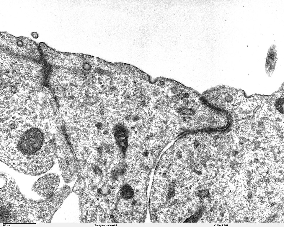
Transmission electron microscope image of a thin section cut through the developing brain tissue (telencephalic hemisphere) of an 11.5 day mouse embryo. This image of the luminal surface of the telencephalon, shows junctional complexes and pinocytotic vesicles. The junctional complex is divided into three types of junctions: 1) the most apical is the tight junction, which controls and/or restricts the movement of molecules across epithelial layers and helps maintain polarity, 2) the zonula adherens, which also includes the numerous actin filaments seen in the apical cytoplasm, and 3) the desmosome, which is a spot junction. The pinocytotic vesicles are formed from coated pits in the plasma membrane and are involved in endocytosis. JEOL 100CX TEM References: Marin-Padilla, M. (1985) "Early Vascularization of the Embryonic Cerebral Cortex: Golgi and Electron Microscope Studies", J. Comparative Neurology, 241:237-249 Marin-Padilla, M. and M. Amievo (1989) "Early Neurogenesis of the Mouse Olfactory Nerve: Golgi and Electron Microscope Studies", J. Comparative Neurology, 288:339-352 |
| Source | |
| Author | Louisa Howard, Miguel Marin-Padilla |
| Permission (Reusing this file) |
PD |
Licensing
| This work has been released into the public domain by its author, Louisa Howard, Miguel Marin-Padilla. This applies worldwide. In some countries this may not be legally possible; if so: Louisa Howard, Miguel Marin-Padilla grants anyone the right to use this work for any purpose, without any conditions, unless such conditions are required by law.
|
File history
Click on a date/time to view the file as it appeared at that time.
| Date/Time | Thumbnail | Dimensions | User | Comment | |
|---|---|---|---|---|---|
| current | 21:06, 2 November 2006 | | 1,600 × 1,278 (861 KB) | Patho | {{Information |Description=Transmission electron microscope image of a thin section cut through the developing brain tissue (telencephalic hemisphere) of an 11.5 day mouse embryo. This higher magnification image of "Embryonic brain 80415", shows an area o |
File usage
The following page uses this file:
Global file usage
The following other wikis use this file:
- Usage on bs.wikipedia.org
- Usage on ca.wikipedia.org
- Usage on de.wikipedia.org
- Usage on de.wikibooks.org
- Usage on et.wikipedia.org
- Usage on fr.wikipedia.org