File:Cilia.jpg
No higher resolution available.
Cilia.jpg (640 × 455 pixels, file size: 75 KB, MIME type: image/jpeg)
|
|
This is a file from the Wikimedia Commons |
Summary
| Description | |
| Source | http://remf.dartmouth.edu/images/ciliaTEM/source/1.html |
| Author | Katherine Connolly - Dartmouth Electron Microscope Facility |
| Permission (Reusing this file) |
http://remf.dartmouth.edu/imagesindex.html |
Licensing
| This work has been released into the public domain by its author, Dartmouth Electron Microscope Facility. This applies worldwide. In some countries this may not be legally possible; if so: Dartmouth Electron Microscope Facility grants anyone the right to use this work for any purpose, without any conditions, unless such conditions are required by law.
|
This file was reviewed on 18:57, 13 October 2011 (UTC) by the administrator or trusted user Common Good (talk), who confirmed the Public Domain status on that date.
|
Original upload log
Originally from en.wikipedia; description page is (was) here
- 19:10, 28 December 2002 Magnus Manske 350x263 (24,575 bytes) (Source and public domain notice at [http://remf.dartmouth.edu/imagesindex.html])
File history
Click on a date/time to view the file as it appeared at that time.
| Date/Time | Thumbnail | Dimensions | User | Comment | |
|---|---|---|---|---|---|
| current | 18:53, 13 October 2011 | | 640 × 455 (75 KB) | Common Good | high res |
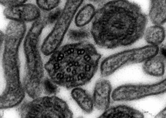
| 19:45, 9 May 2006 |  | 350 × 263 (24 KB) | Magnus Manske | {{Information| |Description= Source and public domain notice at [http://remf.dartmouth.edu/imagesindex.html] TEM image of a thin x-section cut through a en:human nasal en:cilia. Cilia cross |
File usage
The following 2 pages use this file:
Global file usage
The following other wikis use this file:
- Usage on es.wikibooks.org
- Usage on it.wikipedia.org
- Usage on kk.wikipedia.org
- Usage on pl.wikipedia.org
- Usage on pl.wikiquote.org
- Usage on ru.wikipedia.org
- Usage on tr.wikipedia.org
- Usage on uk.wikipedia.org