File:Hair Cell Patterning Defects in the Cochlea.png
Original file (2,012 × 2,475 pixels, file size: 3.05 MB, MIME type: image/png)
|
|
This is a file from the Wikimedia Commons |
Summary
| Description |
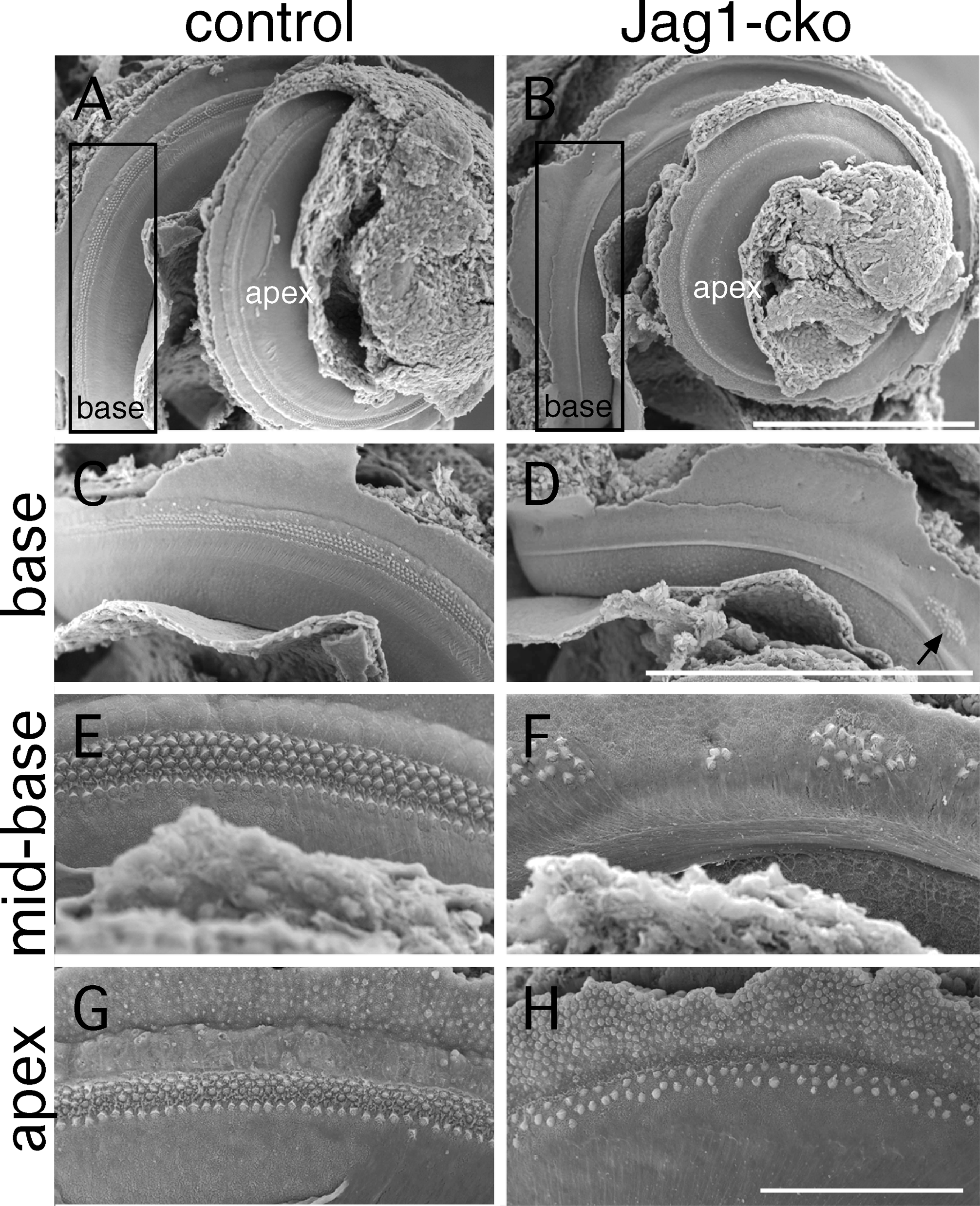
English: Scanning electron micrographs demonstrating the different patterns of hair cell production along the length of the cochlea in Jag1-cko embryos.
(A–D) Low-power views of the apical and basal cochlear turns. The boxed-in area along the base in (A) and (B) is shown at higher magnification in (C) and (D). Note the absence of hair cells in the base of the Jag1-cko cochlea, except for a small patch of cells in the more apical portion (arrow). Scale bars = 500 μm. (E and F) In the midbasal region, more hair cells are observed, but they are arranged in patches, with no clear distinction between inner and outer hair cells. (G and H) In the apical turn, hair cells are continuous but generally arranged in only two rows. Scale bar = 100 μm. |
| Date | |
| Source | The Notch Ligand JAG1 Is Required for Sensory Progenitor Development in the Mammalian Inner Ear ([1]) |
| Author | Amy E. Kiernan, Jingxia Xu, Thomas Gridley |
Licensing
- You are free:
- to share – to copy, distribute and transmit the work
- to remix – to adapt the work
- Under the following conditions:
- attribution – You must give appropriate credit, provide a link to the license, and indicate if changes were made. You may do so in any reasonable manner, but not in any way that suggests the licensor endorses you or your use.
File history
Click on a date/time to view the file as it appeared at that time.
| Date/Time | Thumbnail | Dimensions | User | Comment | |
|---|---|---|---|---|---|
| current | 04:47, 12 February 2009 | | 2,012 × 2,475 (3.05 MB) | Mike.lifeguard | {{Information |Description={{en|1=Scanning electron micrographs demonstrating the different patterns of hair cell production along the length of the cochlea in Jag1-cko embryos. (A–D) Low-power views of the apical and basal cochlear turns. The boxed-in |
File usage
The following page uses this file: